Qibing Liu1,
Na Lin2,
Daorui Yu1,
Song Li1,
Qiaoqin Xu1,
Xianjing Qin1,
Juntao Zeng3 ![]() ,
Yu Zeng1
,
Yu Zeng1
For correspondence:- Juntao Zeng Email: zengjuntaohyfy@sina.com Tel:+8689831350701
Received: 13 October 2015 Accepted: 1 July 2016 Published: 30 August 2016
Citation: Liu Q, Lin N, Yu D, Li S, Xu Q, Qin X, et al. Hypolipidemic effect of aqueous leaf extract of carmona microphylla G Don. Trop J Pharm Res 2016; 15(8):1673-1680 doi: 10.4314/tjpr.v15i8.12
© 2016 The authors.
This is an Open Access article that uses a funding model which does not charge readers or their institutions for access and distributed under the terms of the Creative Commons Attribution License (http://creativecommons.org/licenses/by/4.0) and the Budapest Open Access Initiative (http://www.budapestopenaccessinitiative.org/read), which permit unrestricted use, distribution, and reproduction in any medium, provided the original work is properly credited..
Purpose: To investigate the hypolipidemic effects of the aqueous leaf extract of Carmona microphylla (Lam.) G. Don. (CAE) in vitro and in vivo.
Methods: The lipid-lowering effect of CAE was investigated in oleic acid (OA)-induced steatosis in HepG2 liver cells, as well as in high-fat diet (HFD)- and triton WR-1339 (TRI)-induced hyperlipidemic mice. The levels of intracellular, serum and/or hepatic total cholesterol (TC); triglyceride (TG); low density lipoprotein-cholesterol (LDL-c); high density lipoprotein-cholesterol (HDL-c); hepatic superoxide dismutase (SOD) activity and malondialdehyde (MDA) were determined by oil-red O staining and appropriate kits.
Results: Treatment with CAE inhibited lipid accumulation in HepG2 cells and reduced the elevated levels of serum TC, TG and LDL-c as well as hepatic TC and TG in hyperlipidemic mice induced by HFD. CAE administration also significantly decreased arteriosclerosis index (AI) and LDL-c/HDL-c ratio, but improved oxidative status as revealed by increased hepatic SOD activity and decreased MDA level. The lipid ameliorating and antioxidative effects of CAE (600 mg/kg) were comparable to those of the standard lipid-lowering drug, sivastatin (5 mg/kg).
Conclusion: These results suggest that C. microphylla aqueous extract (CAE) protects against hyperlipidemia induced by HFD in mice and may find therapeutic application in hyperlipidemic patients.
Introduction
Hyperlipidemia, characterized by elevated levels of total serum cholesterol (TC), triglycerides (TG) and low density lipoprotein-cholesterol (LDL-c), is a major risk factor for the development of coronary artery disease and the progression of atherosclerotic lesions [1,2]. High levels of cholesterol, especially LDL-c, may attach to the walls of blood vessels (if the quantity of HDL is insufficient) to form oxidized LDL, which is highly atherogenic and toxic to vascular cells, leading to a variety of serious diseases including atherosclerosis [3,4].
In hyperlipidemic condition, the activities of superoxide dismutase (SOD) and catalase (CAT) are decreased, which leads to pronounced elevation of reactive oxygen species (ROS), such as malondialdehyde (MDA), and causes vascular and tissue damage [5,6]. Although numerous synthetic lipid-lowering drugs, such as statins, fibrates, niacin, and bile acid sequestrants (resins), are available for combating hyperlipidemia, management of this disorder without accompanying side effects remains a major challenge. Therefore, it is necessary to find drugs with lipid lowering and antioxidant activities that produce little or no side effects. Generally, natural products are the preferred option, and a number of herbal medicines from plants and vegetables have been reported to control hyperlipidemia and related complications in patients [7-12].
Carmona microphylla (Lam.) G. Don., commonly known as Ehretia buxifolia Roxb. and also called “Fujian Cha” in China, is widely distributed in the Guangdong, Hainan, and Taiwan provinces of South China, as well as in subtropical areas of southern and south-eastern Asia [13]. The root of C. microphylla has long been used to treat cachexia and venereal infections and is an antidote to food poisoning [14,15]. In the Hainan province of South China, the leaves of C. microphylla have been used as a folk medicine for the treatment of hyperlipidemia and diabetes. However, the pharmacological effects of C. microphylla have not been validated by scientific studies. In the present work, the antihyperlipidemic and antioxidant effects of an aqueous extract of C. microphylla leaves (CAE) were investigated in oleic acid (OA)-induced steatosis in HepG2 liver cells and in high-fat diet (HFD)-induced hyperlipidemic mice.
Methods
Reagents
Triton WR-1339, oleic acid, sivastatin and atorvastatin were purchased from Sigma Chemical Co. (St. Louis, MO, USA). All other chemicals and reagents used were of analytical grade and were obtained from Sinopharm Chemical Reagent Co., Ltd (Beijing, China).
Preparation of C. microphylla leaf extract
Fresh leaves of C. microphylla were collected in May 2013 from Haikou City, Hainan Province, China and were identified by Prof. Niankai Zeng of the College of Pharmacy, Hainan Medical University. A voucher specimen was deposited in the Hainan Medical University herbarium (no. HMU-ETHPH-FJC-1305) for future reference. Freshly harvested leaves were dried and finely powdered. Fifty grams of the powder were extracted with 800 mL of distilled water using a Soxhlet apparatus. The residue thus obtained was then filtered, and the resulting filtrate was concentrated to a dry mass by vacuum distillation; this was used as the C. microphylla aqueous extract (CAE).
Ethics statement
This study was carried out in strict accordance with the recommendations of the Guide for the Care and Use of Laboratory Animals [16]. All procedures with animals were approved by the Institutional Animal Ethical Committee of Hainan Medical University (ref nos. HMU20130135 and HMU20150323).
Experimental animals
Healthy female C57BL/6 mice (18-22 g) were housed under conditions of controlled temperature (25 ± 2 °C) with a 12-h/12-h day-night cycle, during which time, they had free access to food and water ad libitum. The animals were allowed to acclimatize for 5 days before commencing the experiments.
Evaluation of anti-hyperlipidemic effect
HFD-induced hyperlipidemia model
Animals were divided into six groups consisting of eight animals each. The control group was fed with a normal diet, while the other groups were fed with a high fat-diet (HFD). The normal rodent chow contained 12 % fat, 62 % carbohydrate, and 26 % protein, with a total energy content of 12.6 kJ/g. The HFD was formulated to balance micronutrient content on a calorie basis and contained 60 % fat, 26 % carbohydrate, and 14 % protein, with a total energy content of 21.0 kJ/g. The HFD contained much less choline bicitrate (0.6 g/kg) and DL-methionine (1.5 g/kg) than the normal rodent chow. The fatty acid composition of the fats (mainly from lard) in the HFD was 36 % saturated fatty acids, 45 % monounsaturated fatty acids, and 19 % polyunsaturated fatty acids (PUFA). The sivastatin group was treated with 5 mg/kg sivastastin, while the CAE groups were treated with different doses of CAE (10, 50 and 100μg/mL). At the end of the 4-week period, animals were sacrificed after an overnight fast. Blood and liver samples were then collected for the lipid profile and oxidative status.
Triton WR-1339-induced hyperlipidemia model
Animals were divided into five groups, consisting of eight animals each, and treated as follows. A normal control group was treated with vehicle (distilled water), while a TRI control group was given equal volume of distilled water and served as a negative control. The TRI + atorvastatin and TRI + CAE groups were treated with atorvastatin (5 mg/kg body weight) and CAE (300 and 600 mg/kg body weight), respectively. The extract and atorvastatin were administered by oral gavage. After 7 days of treatment, the normal control group was injected with equal volume of distilled water while all other groups were hyperlipidemia-induced by a single intraperitoneal (ip) injection of triton WR-1339 (300 mg/kg body weight) dissolved in normal saline (pH 7.4). Animals were sacrificed 24 h after the injection. Blood was collected directly from the heart of each animal, and the serum was separated and used for the serum lipid profile.
Determination of serum lipid profile
TC, TG, LDL-c and HDL-c were enzymatically determined using commercial kits (BioSino Bio-technology and Science Inc., Beijing, China). Arteriosclerosis index (AI) was calculated as in Eq 1.
AI = (TC-HDL-c)/HDL-c ……………………. (1)
Determination of hepatic antioxidant status
The liver was excised and washed thoroughly in ice-cold saline to remove blood. Ten percent homogenate was prepared in phosphate buffer (0.05 M, pH 7) using a polytron homogenizer. The homogenate was centrifuged at 3000 g for 20 min to remove cell debris and nuclei, and the supernatant was centrifuged at 9000 g for 20 min to remove mitochondria. The supernatant was used for the determination of hepatic levels of MDA, activity of liver SOD, and total protein using a commercial kit (BioSino Bio-technology and Science Inc., Beijing, China).
Cell-based lipid accumulation assay
HepG2 cells were maintained in DMEM (Dulbecco’s Modified Eagle Medium) supplemented with 10 % fetal bovine serum and penicillin/streptomycin (100 μg/mL). The cells with 70 %-80 % confluence were incubated in DMEM + oleic acid (100 μM) for 12 h and were then treated with CAE (as indicated) and the positive control sivastatin (5 μg/mL) in DMEM/100 μM oleic acid, with DMEM/100 μM oleic acid as a blank for an additional 6 h. Subsequently, the cells were subjected to oil-red O staining or TC and TG determination as described previously. Each experiment (n = 8 for oil-red O staining or n = 3 for TC and TG determination) was repeated in triplicate.
Statistical analysis
Data are expressed as the mean ± SEM. One-way analysis of variance (ANOVA) followed by Tukey’s test was used for statistical analysis. SPSS (version 16) statistical software was used for the analysis of data, and p < 0.05 was considered statistically significant.
Results
CAE inhibited lipid accumulation in HepG2 cells
As shown in , supplementation with OA significantly increased lipid accumulation in HepG2 cells. Treatment with CAE decreased OA-elicited neutral lipid accumulation (A,B) as well as the intracellular contents of TC (C) and TG (D) in a dose-dependent manner. This inhibitory effect on lipid metabolism was independent of the cytotoxic effect of CAE on HepG2 cells as revealed by an MTT assay (data not shown).
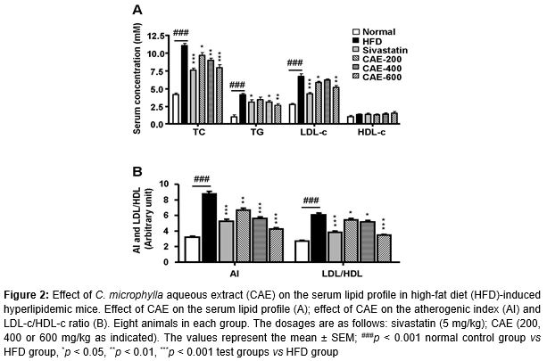
Effect of CAE on the serum lipid profile in high-fat diet (HFD)-induced hyperlipidemic mice
Serum TC, TG and LDL-c levels were significantly elevated (p < 0.001) in the HFD-treated groups (A). Oral administration of CAE to mice resulted in significant decrease of serum TC, TG and LDL-c. The HDL level was increased but not significant when compared to the control group. The lipid-lowering efficacy of CAE at 600 mg/kg was comparable to that of the standard reference drug sivastatin (5 mg/kg) (A). Simultaneously, the atherogenic index (AI) and LDL-c/HDL-c ratio were significantly increased in HFD mice and declined after treatment with CAE for 4 weeks. The efficacy of CAE at 600 mg/kg was also comparable to that of sivastatin (5 mg/kg) (B).
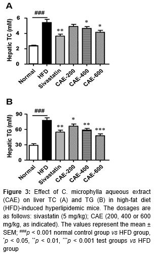
Effect of CAE on hepatic TC and TG levels in HFD-induced hyperlipidemic mice
Consumption of CAE markedly reduced the liver content of TC (5.27 %, 14.14 % and 24.91 % by 200, 400 and 600 mg/kg CAE, respectively) (A) and TG (15.72 %, 28.01 % and 40.11 % by 200, 400 and 600 mg/kg CAE, respectively) (B). At these doses, the efficacy of CAE in decreasing hepatic TC and TG was comparable to that of 5 mg/kg sivastatin, especially in decreasing hepatic TG (B).
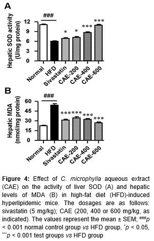
Effect of CAE on the hepatic antioxidant status
The activity of the antioxidant enzyme SOD was significantly lowered while the hepatic level of MDA was significantly increased in the livers of mice treated with HFD (). The CAE treatment improved the activity of SOD and decreased MDA concentration in the liver. The hepatic SOD activity and MDA levels were restored to normal control levels in CAE-treated animals at the dose of 600 mg/kg ().
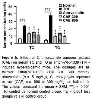
Effect of CAE on serum TC and TG in Triton WR-1339-induced dyslipidemic mice
Serum TC and TG levels were significantly elevated in TRI treated groups (). Treatment with CAE significantly decreased serum TC and TG. The levels of TC and TG were decreased approximately 47.78 % and 49.94 % in the case of 300 mg/kg and 55.96 % and 74.90 % in the case of 600 mg/kg CAE treated groups, respectively, compared to the TRI control (). The standard reference drug atorvastatin (5 mg/kg) showed 53.66 % and 71.00 % decreases for TC and TG, respectively ().
Discussion
The incidence of hyperlipidemia is increasing at a dramatic rate throughout the world [8]. The leaves of Carmona microphylla (Lam.) G. Don. have been used for the treatment of hyperlipidemia in the Hainan province of South China for hundreds of years. However, the pharmacological effects of C. microphylla have not been validated by scientific studies.
In this study, the lipid-lowering effects of C. microphylla aqueous extract (CAE) were first demonstrated in OA-induced steatosis in HepG2 liver cells. Treatment with CAE (100 μg/mL) significantly inhibited lipid accumulation induced by oleic acid in HepG2 cells and largely reduced the intracellular levels of TC and TG with an efficacy comparable to 10 μg/mL of the approved drug sivastatin.
Further evidence was shown in HFD-induced dyslipidemic mice to test the antihyperlipidemic effects of CAE. As is well-known, elevated levels of serum TC, TG and LDL-c, accompanied by reduced HDL-c levels, are frequently associated with an increased risk of coronary heart disease and atherosclerosis [2,17]. The deposition of LDL-c in the blood vessel walls is a major component of atherosclerotic plaque lesions. Moreover, LDL-c is susceptible to oxidative modification, and this oxidative modification of LDL-c is causally involved in the initiation and promotion of atherosclerosis [18]. Hence, decreasing serum TC, TG and LDL-c levels is important for reducing the risk of atherosclerosis.
Treatment with CAE once daily for 4 weeks can effectively decrease the level of serum TC, TG and LDL-c. Hence, through the hypolipidemic effect, this plant extract experimentally supported its efficacy in preventing the early event in atheroma formation. The efficacy of CAE (600 mg/kg) in decreasing serum TC and LDL-c was slightly weaker than sivastatin (5 mg/kg) but more potent in decreasing TG. These results indicate a discrepancy between CAE and statins in lipid-lowering action.
The non-ionic detergent Triton WR-1339 can block the uptake of TG-rich lipoproteins from plasma by peripheral tissues, leading to acute hyperlipidemia in animals. It is widely used, particularly in mice, for screening natural or chemical hypolipidemic drugs [16, 19-20]. In the present study, TC and TG levels were largely elevated 24 h after the triton WR-1339 administration, which was significantly decreased by the treatment with CAE. These findings suggest that the cholesterol-lowering activity of CAE may act through the enhanced catabolism of TC, TG and LDL-c.
HDL carries cholesterol and cholesterol esters from the peripheral tissues and cells to the liver, where cholesterol is metabolized into bile acids. This pathway plays an important role in reducing cholesterol levels in the blood and peripheral tissues and in inhibiting atherosclerotic plaque formation in the aorta [21-22]. The atherogenic index and LDL-c/HDL-c ratio are thus important diagnostic indicators of the risk of atherosclerosis development [23]. In the present study, administration of CAE slightly increased serum HDL-c and significantly decreased TC and LDL-c levels, thus significantly reducing the AI and LDL-c/HDL-c ratio. This ameliorative action provides support for using CAE to prevent atheroma formation. Interestingly, the decrease of the AI and LDL/HDL ratio by CAE at 600 mg/kg was more pronounced than that resulting from sivastatin (5 mg/kg) treatment, indicating a beneficial role for drinking CAE for the prevention of atherosclerosis.
Oxidative stress has been implicated in many diseases including arteriosclerosis. Living tissues are endowed with innate antioxidant defense mechanisms, including the SOD, which is involved in the disposal of superoxides and hydrogen peroxide (H2O2). A reduction in the activity of SOD is associated with the accumulation of highly reactive free radicals, which leads to deleterious effects such as loss of integrity and function of cell membranes [24]. In the present study, the activity of SOD in hepatic tissue samples from hyperlipidemia-induced, distilled water-treated mice was significantly lower than that of normal control mice. Administration of CAE significantly enhanced the activity of SOD in the liver and was more potent than sivastatin and similar to other popular teas.
Lipid peroxidation, a free radical-mediated propagation of oxidative insult to polyunsaturated fatty acids, is a characteristic feature of hyperlipidemia; it impairs cell membrane fluidity and alters the activity of membrane-bound enzymes and receptors, resulting in membrane malfunction [25,26]. The observed elevated mean levels of MDA in HFD control mice possibly resulted from increased intensity of lipid peroxidation, which has been reported to occur from intensified free radical production [27]. Oral administration of CAE to hyperlipidemic mice resulted in significantly lower mean levels of MDA than that of distilled water-treated hyperlipidemic mice. CAE possibly up-regulated hepatic SOD activity, thereby preventing oxidative modification of lipoprotein, thus leading to a reduction in MDA levels.
Conclusion
The present study has demonstrates for the first time the putative lipid-lowering and antioxidant activity of aqueous leaf extract of C. microphylla in oleic acid-elicited HepG2 liver cells and HFD- as well as Triton WR-1339-induced hyperlipidemic mice. The lipid-lowering potential and antioxidant capacity of CAE at a dose of 600 mg/kg appeared to be as effective as that of the standard lipid-lowering drug, simvastatin, at a dose of 5 mg/kg. Hence, CAE can potentially be developed as an alternative cholesterol-lowering drug for clinical use.
Declarations
Acknowledgement
References
Archives


News Updates